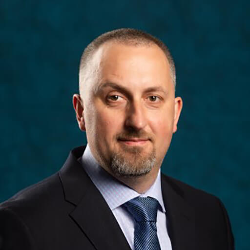
Robert
Zawadzki
PhD
Location
Davis, CA, USA
Current Organization
University of California, Davis
Biography
I am an experimental physicist by training. I began my scientific career researching optical imaging methods in biology and medicine. During my PhD, I worked on the early development and application of Optical Coherence Tomography (OCT). After finishing my PhD, I joined UC Davis, where I started working on combining OCT with Adaptive Optics (AO) and Scanning Light Ophthalmoscopy (SLO) for cellular resolution clinical retinal imaging. Since then, my research focus gradually expanded from the development of high-resolution in vivo imaging modalities to its applications to investigations of cellular resolution morphology and function of the retina and retinal vasculature and its dynamic changes in various types of retinal and optic nerve diseases. Desiring to get a deeper knowledge of the cellular biology of the retina, about eight years ago, I began a collaboration with Prof. Edward N. Pugh Jr. and created a shared mouse ocular imaging facility, the “EyePod”. I have a broad background in biomedical optics, biomedical engineering, and vision science. I have recently co-developed a noninvasive Temporal Speckle Analysis OCT (TSA-OCT) method, employing dedicated image analysis software, allowing imaging of RGC structures at microscopic (somas and axons) and macroscopic (GCL and RNFL) levels with corresponding functional assessment of dynamic scattering. Thus, allowing direct comparison and validation of the predictive value of TSA-OCT for individual RGCs fate in Glaucoma.


